General discussion
This is chapter 6 of my PhD thesis, defended at Radboud University in . It was written for the thesis and has not been published elsewhere.
Overview
In this thesis, we set out to improve prostate cancer diagnostics by applying artificial intelligence (AI). With the goal to bring these innovations to the clinic, we cannot discuss AI systems independently of the target domain. Namely, the autonomy of an AI system, its design choices, and its impact on the physician’s workflow vary according to its intended use (Figure 6.1). For example, a system could take a prioritization role and filter biopsies before the pathologist’s review. A large portion of evaluated prostate biopsies does not contain cancer1 and pathologists’ workload can be drastically reduced if an AI system filters these out. Filtering on its own does not improve the quality of the diagnosis, though. Additionally, the high autonomy of prioritization systems, especially if it screens biopsies, will require rigorous testing to prevent missing clinically relevant cancers.
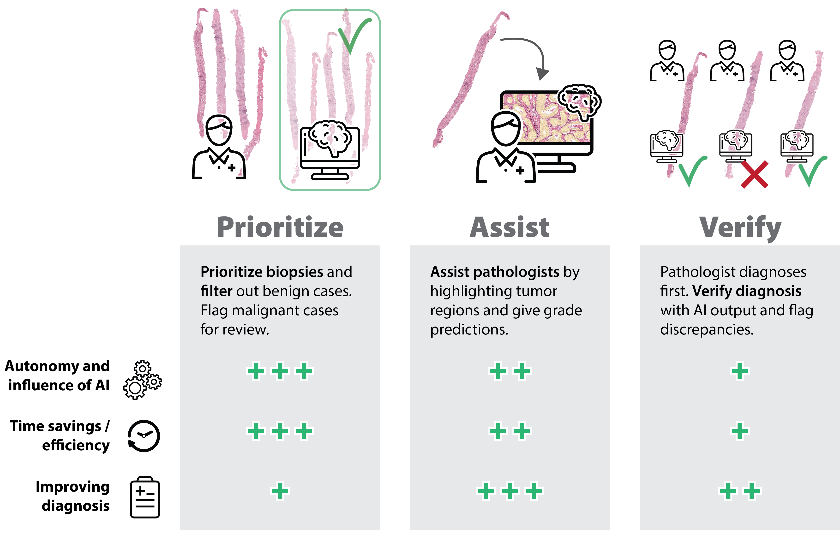
Mirrored to the screening approach is an AI system that verifies the pathologists’ diagnosis. Unfortunately, this method results in almost zero time gains as the pathologist still performs a regular review and could even reduce efficiency, as more cases will get a second read. Nevertheless, it is a promising approach to increase overall sensitivity, though one should investigate whether this would result in overtreatment of non-significant cancers.
Besides screening and verifying, an AI system can also actively support pathologists during the diagnostic process. This approach is a middle ground in terms of autonomy and influence: The AI system is actively involved, but a pathologist can always override the predictions. By combining the AI system’s and pathologist’s expertise, there is a high potential for improving the quality of the diagnosis. Time-wise, some improvements can be expected as the AI can pre-fill reports and guide pathologists, reducing time spent searching for relevant tissue regions. Overall this AI assistance can be seen as an interactive second pair of eyes: a digital fellow.
The investigation and design of such a ‘digital colleague’ was the main focus of this thesis. Specifically, we aimed to support pathologists using deep learning-based methods when grading prostate biopsies following the Gleason grading system. In this final chapter, we evaluate our work, assess the impact made on the field, and outline future directions.
Fine-grained segmentations
At the start of this research, automated histological assessment of prostate cancer was still in its infancy – with still a significant focus on feature engineering24 – but the field was transitioning quickly to deep learning-based methods. While promising as a technique, these existing methods were often very coarse in their output, limiting their application.5 This coarseness was primarily present in these model’s inability to distinguish between epithelial and stromal tissue, often caused by a lack of detailed training data. Algorithms that could segment with higher precision were desired for correct quantification of tumor areas or for displaying results to pathologists.
Acquiring large amounts of highly detailed data was, and still is, a challenge within computational pathology. Unique to pathology is the ability to use different staining techniques to highlight certain cell types or structures in the tissue. Chapter 2 of this thesis aimed to overcome the annotation burden by employing immunohistochemistry (IHC) to annotate data for model training semi-automatically. Using CK8/18 and P63 to highlight the epithelial tissue and basal cell layer, we could accurately distinguish between the epithelial and stromal tissue. Restaining and registering slides allowed us to map the semi-automatic annotations made using the IHC slides to the H&E versions. This technique showed to speed up data labeling significantly and removed the subjectiveness associated with human-made annotations. Due to this IHC-backed reference standard, the final deep learning model was able to segment the epithelial tissue with higher precision than reasonably achievable with just human annotations. The dataset was released publicly for research use as the PESO datsaset.6
On its own, a segmentation algorithm for epithelial tissue is of little clinical use. However, the ability to accurately segment epithelial tissue can drastically reduce the annotation burden for higher-level annotation tasks, e.g., cancerous glands. Additionally, the techniques we showed in this chapter are not limited to epithelium segmentation. On the contrary, using IHC to segment tissue semi-automatically is an effective strategy to create detailed annotations and applicable to many tasks within computation pathology.
AI-powered Gleason grading
For patients with prostate cancer, the Gleason score and grade group of a biopsy are among the most important prognostic markers.79 Unfortunately, the grading system suffers from substantial inter- and intraobserver variability,1012 and while specialized uropathologists show a higher agreement,13 they are not available to every patient. At this point, most existing studies were done on preselected, smaller subimages or tissue microarrays.241417 Studies that focussed on full whole slide images either used a small dataset with a subset of Gleason patterns18 or prostatectomies.19
Chapter 3 investigated whether a deep learning system could grade prostate biopsies at expert levels to make high-quality reproducible Gleason grading more accessible to patients. A substantial part of this project consisted of compiling a new cohort of 5759 prostate biopsies as there was no pre-existing annotated public dataset for Gleason grading available. Annotating this new dataset would quickly require hundreds of pathologist hours. To remedy this, we used a novel pipeline to semi-automatically annotate the data on a gland-by-gland level using the developed epithelium segmentation system (Chapter 2) and an existing tumor detection system.5
The subjective nature of Gleason grading also introduced challenges when assessing the algorithm’s performance. The gold standard for reference standards consists of asking multiple specialized uropathologists to grade biopsies and combining their opinions. Still, while using a panel improves the reference standard’s quality, it is not watertight and remains dependent on a correct assessment by pathologists. It also limits the upper bound of a system’s performance to the experts who set the reference standard. When algorithms improve in performance, we will eventually reach a point where we can argue who is right.
To determine the added value of automated grading for clinical practice, we assessed whether the new technique is non-inferior – and ideally better – compared to the standard practice of care. Thus, we compared the AI system with the grading performance of a panel of pathologists and residents. We found that, on average, the AI system showed a higher agreement with the experts than the panel, operating at the level of experienced pathologists, and giving concrete proof of the power of this technology.
Several other studies have confirmed our findings and demonstrated the high performance of deep learning-based Gleason grading of biopsies.1922 Most notably are the works by Ström et al.,21 published simultaneously in the same journal,23 and the later work by Nagpal et al.22 In output and approach, these studies were quite similar to ours, both used deep learning-based models albeit with different architectures. The main differences were twofold: We used an annotation-less approach of collecting the data based on the diagnosis of the original pathologist report and without extensive annotations by pathologists. Secondly, we determined the specimen’s grade group in the same way as pathologists would do in clinical practice, instead of using an additional machine learning model. Thus, as our model detects Gleason patterns on a glandular level, we can directly calculate the primary and secondary patterns and determine the grade group. This technique ensures that the model output is easily interpretable and there is no black box that determines the final diagnosis.
The PANDA Challenge
Chapter 3 showed the promise for accurate AI-based diagnosis and grading of prostate cancer in biopsies. However, despite our results and that of similar studies,212224 there had been limited proof of generalization to diverse and multinational patient cohorts and reference standards. Additionally, these studies – as common in the field – were developed in a siloed matter: the same researchers who developed the algorithms also validated them. Thus, to assess the maturity of AI for Gleason grading, we required a more independent validation of these new techniques.
Medical imaging competitions, or challenges, have shown great accelerators for AI innovations, with the CAMELYON challenges being a supreme example.2526 However, many of these competitions were also limited in their independent validation and reproducibility of algorithms, raising doubt whether competitions solve the underlying medical problem.27
We aimed to address both issues in Chapter 4 through organizing the PANDA challenge: an international competition on AI-based Gleason grading. With a development set of 10,616 biopsies, 2,009 biopsies for internal and external validation, and 1,290 participants, it was the largest competition organized for pathology to date.28 We required that all challenge participants submitted working algorithms, which were validated entirely independently of the algorithm developers, reducing sources of potential bias and verifying the reproducibility of the proposed solutions. While this approach requires significantly more compute resources to organize challenges, it is a logical evolution from previous challenge designs and should become the defacto standard in the field. Open research platforms such as grand-challenge.org could play a significant role in this evolvement of challenge designs by making these study setups more accessible.
As all algorithms from PANDA were reproducible, it allowed us to apply algorithms to external cohorts and to assess AI’s performance for Gleason grading as a ‘whole’ instead of individual algorithms. Evaluating a group of algorithms is distinctly different from evaluating just the ‘best’ algorithm of a competition, which can suffer from limitations and biases.27 We found that algorithm performance was retained even on cross-continental datasets, showing that algorithms trained on EU data can generalize to US-based cohorts. Algorithms were highly sensitive for cancer, missing fewer cancers than pathologists, but suffered in terms of specificity on the external validation sets.
Organizing a competition in this way, ensuring independent validation and reproducibility, adds significant evidence of the maturity of AI for automated Gleason grading. In addition, it is one of the most extensive evaluations that can be done within the limits of a retrospective setup. The next logical step is to validate these methods in clinical practice, e.g., by allowing pathologists to use these technologies and through prospective clinical trials.
The added value of AI-assistance
AI successes for medical applications co-occur with a lot of hype and fear that AI could make physicians obsolete.2932 While the diagnostic power of these AI systems is not generally contested, there is still a gap between research results and clinical practice. Even though a system can achieve a high agreement with experts or accurately outline a tumor region, this does not directly translate to improvements in the diagnostic process or better care.
In the research community, there is disproportionate attention for achieving state-of-the-art (SOTA) results: ample new methods are being introduced that claim to achieve SOTA on benchmark datasets. As such results make for good headlines, the reasoning is clear, and of course, we want to drive the field forward to achieve optimum performance. However, it is sometimes questionable whether these slight increases in performance actually result from better algorithm design; It is one reason we opted for a group-based analysis for the PANDA challenge instead of focusing solely on the challenge winner.
To truly evaluate algorithms’ added benefits, we need to assess them in a suitable environment and investigate how we could implement such systems. These evaluations go beyond quality metrics and achieving top scores on benchmarks and should include efficiency assessments and human-computer interaction as well.33
When we completed our work on automated grading (Chapter 3), there were countless other papers on AI for pathology. However, studies showing how these techniques can be used effectively were still rare. There was limited evidence of the merits of AI assistance for the histological assessment of prostate cancer. Some studies have been performed to show the value of AI assistance in detecting prostate cancer,34 but no such study existed for Gleason grading. In Chapter 5, we showed that pathologists could actually benefit from AI assistance when diagnosing prostate cancer.
In Chapter 3, the AI system outperformed most pathologists, but the AI-supported pathologists now outperformed both the standalone AI as the pathologists without AI. This manifestation of pathologist-AI synergy can be easily explained as both the pathologist and AI system have different strengths and weaknesses. AI systems are consistent, reproducible, and cannot get tired. As a result, all tissue is analyzed consistently, and there is a lower chance that small tumors are missed. Additionally, AI systems offer high precision in quantification, allowing pathologists to better assess the tumor’s extent. On the other hand, algorithms can notoriously fail in the presence of data patterns that were not in the training dataset; examples include artifacts, foreign tissue, or extensive shifts in the stain characteristics. Pathologists will more easily adjust to changes in the staining characteristics and disregard artifacts.
To the best of our knowledge, we were the first to show that AI-assisted can help pathologists perform better at Gleason grading. Shortly after, Steiner et al. also demonstrated the benefits of AI assistance for Gleason grading on a different cohort.35 These results are fundamental for the field; we not only need to show the power of AI innovations but more so proof that they can be effectively used in practice. Following this, as was already said for radiologists,31 one could say that AI will not directly replace pathologists, but pathologists using AI could replace those who do not.
On the importance of data
An alternative fitting title of this work could have been “improving prostate cancer grading using big data.” While technological advances in AI, and specifically deep learning, have allowed researchers to achieve state-of-the-art results, the access to large datasets continues to fuel this revolution. In contrast to the old way of explicit feature engineering, current AI applications for medical imaging regularly use existing architectures or a combination of existing building blocks. The availability of large datasets, combined with the tools and computing power to harness those, forms the basis of many recent advancements. Additionally, there is a positive correlation between dataset size and model performance.36 The chapters of this thesis show a similar trend, from 102 cases (Chapter 2) to a cohort of more than 11,000 biopsies (Chapter 4), and can also be seen in many other studies which consistently include large datasets.1921223739
With data at the core of AI development, the collection, curation, and intelligent processing of those datasets will only become more and more critical. The results presented in this thesis would not have been possible without novel ways of collecting and transforming the data for model training (Chapter 2 and 3). Even though there is much attention for new methodologies, for many of the current pathology tasks, AI development is probably best started with a good grasp of the data, with the actual deep learning architecture or method following second.
Unfortunately, while there is typically fast dissemination of new methodological ideas, the same does not hold for data. Datasets, especially in the medical domain, are often well guarded by the institutions that collected them. Most of this is understandable, either from privacy perspectives or because of the inherent value data holds for research groups. However, because data is so vital, it can also limit some of the high-impact applied research to those who can afford it. By publishing two unique datasets for computational pathology, the PESO6 and PANDA40 datasets, we aimed to contribute to the availability of research data and fuel further innovations. Despite that these datasets are new, they have already been downloaded thousands of times. The PESO dataset was used in several internal244142 and external publications,4345 and we expect considerable interest in the PANDA dataset due to its size and application.
Between now and clinical practice
The majority of this thesis has focussed on quantitative analysis of performance, either that of automated systems or AI-assisted pathologists. However, the actual implementation of these techniques goes beyond achieving high performance on independent validation cohorts.4647 With higher autonomy and decision-power of AI systems, we also need to focus on the implementation and robustness of such systems, especially from an end-user perspective.
To start, most of the challenges introduced in Chapter 1 regarding scanner and stain variations remain highly relevant. An algorithm can work flawlessly on a test cohort that originated from the same center as the training cohort, but for real-world scenario’s we need to assure quality among a wide range of image characteristics. Applying techniques such as data augmentation to increase the diversity of the training data and (deep learning-based) normalization can help algorithms become more robust.414849 However, these techniques also have their limits, and building multi-center multi-scanner datasets will remain essential in tackling these problems.
With measures to increase robustness in place, it is still not guaranteed that algorithms will be able to handle all input data. Therefore, it would be sensible to require that quality-assurance (QA) algorithms precede clinical algorithms. Not only from a safety aspect but also to not waste pathologists’ time with faulty predictions due to the garbage-in-garbage-out principle. Such algorithms could check for artifacts, ensure that the tissue is the right type for the clinical algorithm, determine if the stain is of sufficient quality, no tissue is out of focus, and outline valid regions where the clinical algorithm should be applied. Utilizing these techniques, combined with on-site validations, will be vital to retaining model performance across clinics and ensure stable day-to-day operations.
Beyond increasing resilience to data variants and artifacts, we need actual fail-safes that prevent incidents from going unnoticed. Unlike their human colleagues, AI systems in their default forms are often limited in expressing uncertainty and often suffer from catastrophic failure when confronted with data outside the training distribution.5051 Commonly, samples that are not within the set of known classes are still classified as one of those and with high confidence values. When AI is applied outside of the strictly controlled research settings, we will need tools to express certainty, detect these outliers, and flag cases accordingly.5254 Even if a QA algorithm accepted a case for further review, the actual medical device should still alert the pathologist if a diagnosis cannot be made with certainty. The pathologist that uses the AI can then determine not to take the AI’s predictions into account. This out-of-distribution detection and uncertainty estimation is an active field of research and a fundamental ingredient for future AI implementations.
Besides the technical challenges, we should not underestimate the human component when applying AI in clinical practice. In Chapter 5, pathologists used the developed Gleason grading system without any prior training. With proper training and a better understanding of the strengths and weaknesses of an AI system, pathologists will be better equipped to utilize AI in clinical practice.3355 On the other side, we can also adjust how algorithms display predictions to better match pathologists. For example, we can show results in numeric formats, overlays on the tissue, or annotations and outlined regions. The explainability of an algorithm is critical for a clinician’s ability to put the predictions in context56 and visualizations can benefit or harm this. Unfortunately, in research, these more qualitative results on usability and algorithm output are often hidden in the supplemental or not even focussed on at all. Future research in human-computer interaction, or maybe better described as human-algorithm interaction, should guide us in designing systems to match the pathologists that will use them.
To summarize, AI implementation in the pathologist’s workflow co-occurs with many challenges, both technical and from a clinician’s perspective, and not to mention societal issues and regulatory requirements. For Gleason grading specifically, the results of this thesis and related work1921223539 show that the technical foundation is present, but we also have not yet reached clinical practice. Thus, it is time for prospective validation of these innovations, where we investigate what impact AI can have on clinical diagnostics in real-life scenarios. In such a prospective setting, we can then compare AI-based grading to routine diagnostics, investigate its impact on efficiency, and eventually determine the added benefit for patients. In this “last mile” gap between AI research and clinical implementation,46 our challenges will predominantly be focussed on everything but the actual algorithm itself.
Beyond human reference standards
This thesis has focussed primarily on improving and optimizing the current histopathological diagnostics for prostate cancer. The Gleason grading standard has been around for years, and by adding AI assistance, we aim to improve the diagnostic process through reproducibility, higher quality, and increased efficiency. While these are giant steps and could significantly benefit patients, a cynic could also argue that these are mere ‘process optimizations.’ So what does the future hold for (prostate cancer) diagnostics?
The Gleason system was built around the idea that specific tissue patterns correspond to distinct patient survival stratification.57 Humans are good at pattern recognition in a general sense, but it is out of the question that AI can do this at an unprecedented scale and detail. Therefore, the next step forward would be extending or further refining the grading system. Automated quantifications could make grading more reproducible and precise,192158 allowing for more fine-grained updates to the Gleason grading system. In addition, utilizing the power of deep learning models to recognize patterns could give further insight into the many variants and subtleties of prostate cancer.59
Detected patterns are not always practical, though; an AI system can also react to specific image characteristics or implicit biases in the data. E.g., suppose images with high-grade cancer originate from a single institution. In that case, the AI system could learn to associate that institution with high-grade tumors instead of using actual tissue patterns for the classification. Evaluation of such systems is therefore not trivial. When we assess AI systems’ performances on tasks using human-based reference standards, we are ultimately limited by humans’ ability for pattern recognition. Given the high observer variability of Gleason grading, we will reach this limitation fairly quickly.
A transition has been started in the field that moves away from human-defined subjective reference standards (e.g., a Gleason score) to patient-level ground truths.60 For example, using patient survival data as the ground truth could remove the subjectiveness introduced by, e.g., the Gleason score (Figure 6.2). Techniques such as neural image compression,61 streaming CNNs6263 and multiple instance learning3964 allow for model training using solely slide or patient-level labels. As part of the PANDA challenge, many teams already demonstrated that training directly using slide-level labels was an effective strategy, albeit pathologists still supplied these labels.
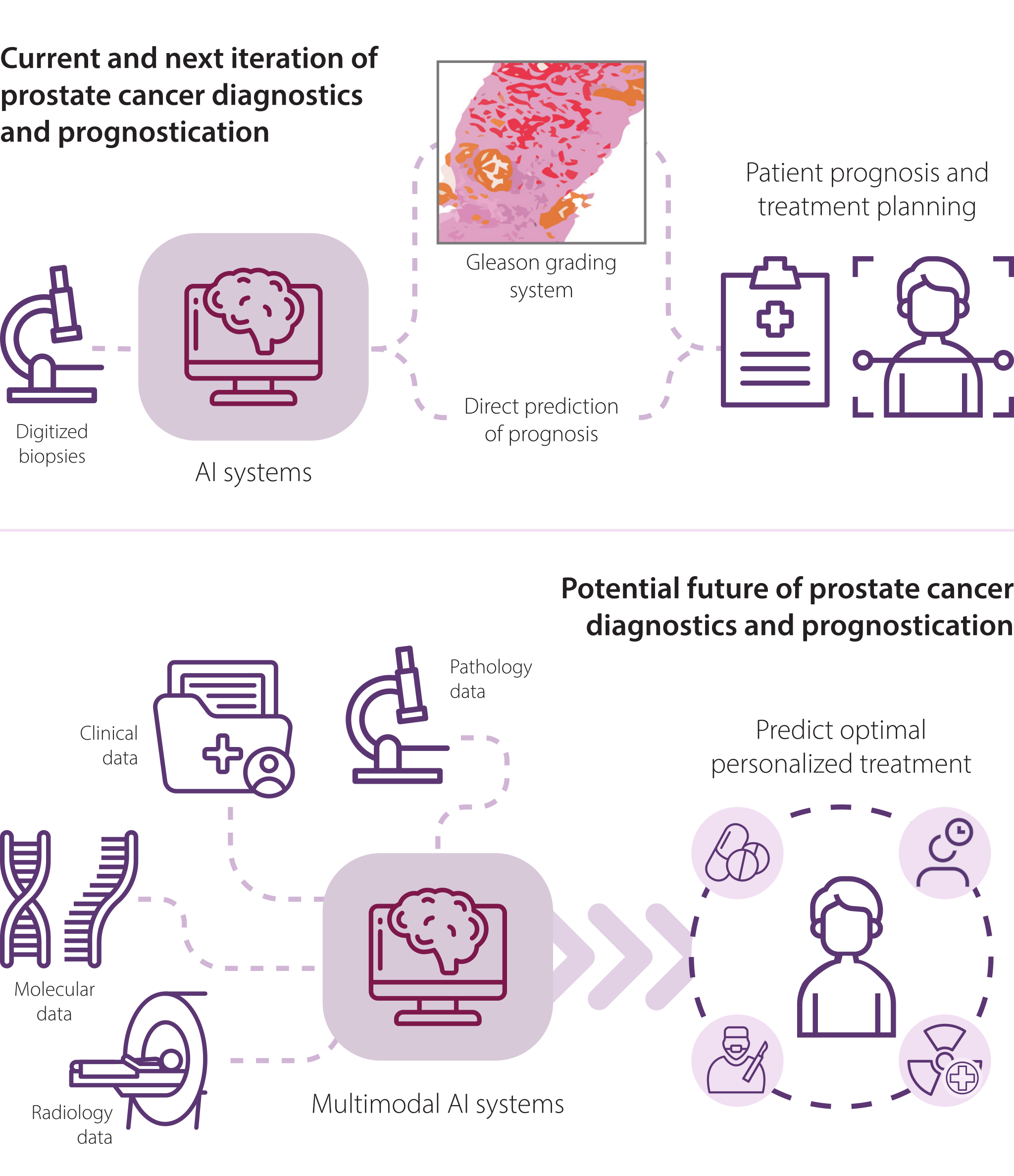
In the future, we will have algorithms that, based on the raw image data, make direct predictions on patient prognosis without any human-defined intermediate. Such digital biomarkers could be the second wave of the AI-powered revolution of clinical diagnostics in pathology. However, defining studies to develop such systems is non-trivial, primarily when based on retrospectively collected data.6566 If we train AI systems on long-term retrospectively collected survival data, the treatment choices based on the pathologist’s original diagnosis could introduce bias and reduce the power of such systems in a prospective setting.
Besides the risk of bias, the second concern of high-level end-to-end systems would be the lack of explainability of such systems. As a result, there is a risk that AI systems could become a black box, even more than some would suggest it is now. A system that detects Gleason patterns and uses this to determine a diagnosis is more investigable than solely a patient-level risk assessment. However, if the black box is validated, tested for bias, achieves high accuracy, and has fail-safes in place, it would benefit the patient to embrace such technology. Adding explainability could therefore be a reverse process. Alternatively to the approach of the Gleason grading system, we will (slowly) move away from manually finding patterns and linking these to prognosis. Instead, an algorithm can directly predict prognosis, and we can try to reverse engineer which patterns contributed to this prediction to allow for human investigation and explainability.6769
Looking beyond the current generation of AI systems and those that predict slide-level labels is a generation of multimodal AI systems (Figure 6.2). Treatment planning for prostate cancer patients, e.g., by a urologist, is not based solely on the Gleason score. Instead, when determining an individual patient’s optimal treatment, a physician will take all available information into account, including data from pathology, radiology, etc. The next generation of AI systems could aggregate all this information, breaking barriers between modalities and hospital departments to determine an individual patient’s optimal personalized treatment plan. This is far from trivial and is not expected to become standard of care soon, but it holds potential for further improving diagnostics and treatment planning.
Conclusion
This thesis described the development and validation of AI-assisted Gleason grading in prostate biopsies. Our work started from the most basic level, finding epithelial tissue in prostate specimens (Chapter 2). We then showed that an AI algorithm achieved pathologist-level performance in a large-scale diagnostic study (Chapter 3). Next, by organizing the most extensive medical image challenge for pathology to date, the PANDA Challenge, we demonstrated that AI for Gleason grading is a mature and promising technology that warrants further validation in prospective settings (Chapter 4). Finally, and perhaps most importantly, we were able to show that AI can indeed improve Gleason grading of prostate biopsies by pathologists (Chapter 5).
The future will hold at least two important directions. First, existing methods can be extended to increase clinical acceptance and streamline implementation, including interpretability, robustness, and interaction with pathologists. These are primarily iterative improvements and not scientific breakthroughs. Still, they are crucial if we want to design AI that actually reaches clinical practice and benefits patients. Second, new AI technologies will allow for novel biomarkers beyond human reference standards and what we use in diagnostics today. Thus, the future is bright for AI-based diagnostics for prostate cancer in pathology, not only from the research and technical perspective but also for pathologists who will harness these new technologies and improve the care they deliver for patients.
References
- Welch HG, Fisher ES, Gottlieb DJ, Barry MJ. Detection of prostate cancer via biopsy in the Medicare–SEER population during the PSA era. Journal of the National Cancer Institute. 2007;99(18):1395–1400.
- Naik S, Doyle S, Feldman M, Tomaszewski J, Madabhushi A. Gland Segmentation and Computerized Gleason Grading of Prostate Histology by Integrating Low- , High-level and Domain Specific Information. In: Proceedings of 2nd Workshop on Microsopic Image Analysis with Applications in Biology. 2007:1–8.
- Gertych A, Ing N, Ma Z, Fuchs TJ, Salman S, Mohanty S, et al. Machine learning approaches to analyze histological images of tissues from radical prostatectomies. Computerized Medical Imaging and Graphics. 2015;46 Pt 2:197–208. doi:10.1016/j.compmedimag.2015.08.002
- Nguyen TH, Sridharan S, Macias V, Kajdacsy-Balla A, Melamed J, Do MN, et al. Automatic Gleason grading of prostate cancer using quantitative phase imaging and machine learning. Journal of Biomedical Optics. 2017;22(3):036015. doi:10.1117/1.jbo.22.3.036015
- Litjens G, Sánchez CI, Timofeeva N, Hermsen M, Nagtegaal I, Kovacs I, et al. Deep learning as a tool for increased accuracy and efficiency of histopathological diagnosis. Nature Scientific Reports. 2016;6:26286. doi:10.1038/srep26286
- Bulten W, Bándi P, Hoven J, Loo RVD, Lotz J, Weiss N, et al. PESO: Prostate Epithelium Segmentation on H&E-stained prostatectomy whole slide images. 2018. doi:10.5281/zenodo.1485967
- Epstein JI. An update of the Gleason grading system. Journal of Urology. 2010;183:433–440. doi:10.1016/j.juro.2009.10.046
- Mohler JL, Antonarakis ES, Armstrong AJ, D’Amico AV, Davis BJ, Dorff T, et al. Prostate Cancer, Version 2.2019, NCCN Clinical Practice Guidelines in Oncology. Journal of the National Comprehensive Cancer Network J Natl Compr Canc Netw. 2019;17(5):479–505. https://jnccn.org/view/journals/jnccn/17/5/article-p479.xml
- Federatie van Medisch Specialisten. Prostaatcarcinoom Richtlijn - Gleason score. Accessed February 26, 2021. https://richtlijnendatabase.nl/richtlijn/prostaatcarcinoom/pathologisch_onderzoek/gleason_score.html
- Allsbrook WC, Mangold KA, Johnson MH, Lane RB, Lane CG, Epstein JI. Interobserver reproducibility of Gleason grading of prostatic carcinoma: General pathologists. Human Pathology. 2001;32(1):81–88. doi:10.1053/hupa.2001.21135
- Egevad L, Ahmad AS, Algaba F, Berney DM, Boccon-Gibod L, Compérat E, et al. Standardization of Gleason grading among 337 European pathologists. Histopathology. 2013;62(2):247–256. doi:10.1111/his.12008
- Melia J, Moseley R, Ball R, Griffiths D, Grigor K, Harnden P, et al. A UK-based investigation of inter-and intra-observer reproducibility of Gleason grading of prostatic biopsies. Histopathology. 2006;48(6):644–654.
- Allsbrook WC, Mangold KA, Johnson MH, Lane RB, Lane CG, Amin MB, et al. Interobserver reproducibility of Gleason grading of prostatic carcinoma: Urologic pathologists. Human Pathology. 2001;32(1):74–80. doi:10.1053/hupa.2001.21134
- Jafari-Khouzani K, Soltanian-Zadeh H. Multiwavelet grading of pathological images of prostate. IEEE Transactions on Biomedical Engineering. 2003;50(6):697–704.
- Källén H, Molin J, Heyden A, Lundström C, Åström K. Towards grading gleason score using generically trained deep convolutional neural networks. In: 2016 IEEE 13th International Symposium on Biomedical Imaging (ISBI). IEEE; 2016:1163–1167.
- Gummeson A, Arvidsson I, Ohlsson M, Overgaard NC, Krzyzanowska A, Heyden A, et al. Automatic Gleason grading of H and E stained microscopic prostate images using deep convolutional neural networks. In: Medical Imaging 2017: Digital Pathology. Vol 10140. International Society for Optics and Photonics; 2017:101400S.
- Arvaniti E, Fricker KS, Moret M, Rupp N, Hermanns T, Fankhauser C, et al. Automated Gleason grading of prostate cancer tissue microarrays via deep learning. Scientific Reports. 2018;8(1):1–11. doi:10.1038/s41598-018-30535-1
- Lucas M, Jansen I, Savci-Heijink CD, Meijer SL, de Boer OJ, van Leeuwen TG, et al. Deep learning for automatic Gleason pattern classification for grade group determination of prostate biopsies. Virchows Archiv. 2019. doi:10.1007/s00428-019-02577-x
- Nagpal K, Foote D, Liu Y, Chen PHC, Wulczyn E, Tan F, et al. Development and validation of a deep learning algorithm for improving Gleason scoring of prostate cancer. npj Digital Medicine. 2019;2(1):48. doi:10.1038/s41746-019-0112-2
- Ryu HS, Jin MS, Park JH, Lee S, Cho J, Oh S, et al. Automated Gleason Scoring and Tumor Quantification in Prostate Core Needle Biopsy Images Using Deep Neural Networks and Its Comparison with Pathologist-Based Assessment. Cancers. 2019;11(12). doi:10.3390/cancers11121860
- Ström P, Kartasalo K, Olsson H, Solorzano L, Delahunt B, Berney DM, et al. Artificial intelligence for diagnosis and grading of prostate cancer in biopsies: a population-based, diagnostic study. The Lancet Oncology. 2020;21(2):222–232.
- Nagpal K, Foote D, Tan F, Liu Y, Chen PHC, Steiner DF, et al. Development and validation of a deep learning algorithm for Gleason grading of prostate cancer from biopsy specimens. JAMA oncology. 2020;6(9):1372–1380.
- Madabhushi A, Feldman MD, Leo P. Deep-learning approaches for Gleason grading of prostate biopsies. The Lancet Oncology. 2020;21(2):187–189.
- Bulten W, Pinckaers H, van Boven H, Vink R, de Bel T, van Ginneken B, et al. Automated deep-learning system for Gleason grading of prostate cancer using biopsies: a diagnostic study. The Lancet Oncology. 2020;21(2):233–241. doi:10.1016/S1470-2045(19)30739-9
- Ehteshami Bejnordi B, Veta M, van Diest PJ, van Ginneken B, Karssemeijer N, Litjens G, et al. Diagnostic Assessment of Deep Learning Algorithms for Detection of Lymph Node Metastases in Women With Breast Cancer. Journal of the American Medical Association. 2017;318(22):2199–2210. doi:10.1001/jama.2017.14585
- Bándi P, Geessink O, Manson Q, van Dijk M, Balkenhol M, Hermsen M, et al. From detection of individual metastases to classification of lymph node status at the patient level: the CAMELYON17 challenge. IEEE Transactions on Medical Imaging. 2018;38(2):550–560. doi:10.1109/tmi.2018.2867350
- Maier-Hein L, Eisenmann M, Reinke A, Onogur S, Stankovic M, Scholz P, et al. Why rankings of biomedical image analysis competitions should be interpreted with care. Nature communications. 2018;9(1):1–13.
- Hartman DJ, Van Der Laak JA, Gurcan MN, Pantanowitz L. Value of public challenges for the development of pathology deep learning algorithms. Journal of pathology informatics. 2020;11.
- Goldhahn J, Rampton V, Spinas GA. Could artificial intelligence make doctors obsolete? BMJ. 2018;363. doi:10.1136/bmj.k4563
- Krittanawong C. The rise of artificial intelligence and the uncertain future for physicians. European Journal of Internal Medicine. 2018;48:e13–e14. doi:10.1016/j.ejim.2017.06.017
- Langlotz CP. Will Artificial Intelligence Replace Radiologists? Radiology: Artificial Intelligence. 2019;1(3):e190058. doi:10.1148/ryai.2019190058
- Ahuja AS. The impact of artificial intelligence in medicine on the future role of the physician. PeerJ. 2019;7:e7702.
- Cai CJ, Winter S, Steiner D, Wilcox L, Terry M. " Hello AI": Uncovering the Onboarding Needs of Medical Practitioners for Human-AI Collaborative Decision-Making. Proceedings of the ACM on Human-computer Interaction. 2019;3(CSCW):1–24.
- Raciti P, Sue J, Ceballos R, Godrich R, Kunz JD, Kapur S, et al. Novel artificial intelligence system increases the detection of prostate cancer in whole slide images of core needle biopsies. Modern Pathology. 2020:1–9.
- Steiner DF, Nagpal K, Sayres R, Foote DJ, Wedin BD, Pearce A, et al. Evaluation of the Use of Combined Artificial Intelligence and Pathologist Assessment to Review and Grade Prostate Biopsies. JAMA Network Open. 2020;3(11):e2023267–e2023267. doi:10.1001/jamanetworkopen.2020.23267
- Sun C, Shrivastava A, Singh S, Gupta A. Revisiting Unreasonable Effectiveness of Data in Deep Learning Era. In: 2017 IEEE International Conference on Computer Vision (ICCV). 2017:843–852. doi:10.1109/ICCV.2017.97
- Gulshan V, Peng L, Coram M, Stumpe MC, Wu D, Narayanaswamy A, et al. Development and Validation of a Deep Learning Algorithm for Detection of Diabetic Retinopathy in Retinal Fundus Photographs. JAMA. 2016;316(22):2402–2410. doi:10.1001/jama.2016.17216
- Ardila D, Kiraly AP, Bharadwaj S, Choi B, Reicher JJ, Peng L, et al. End-to-end lung cancer screening with three-dimensional deep learning on low-dose chest computed tomography. Nature Medicine. 2019;25(6):954–961. doi:10.1038/s41591-019-0447-x
- Campanella G, Hanna MG, Geneslaw L, Miraflor A, Werneck Krauss Silva V, Busam KJ, et al. Clinical-grade computational pathology using weakly supervised deep learning on whole slide images. Nature Medicine. 2019. doi:10.1038/s41591-019-0508-1
- Bulten W, Litjens G, Pinckaers H, Ström P, Eklund M, Kartasalo K, et al. The PANDA challenge: Prostate cANcer graDe Assessment using the Gleason grading system. Zenodo. 2020. doi:10.5281/zenodo.3715938
- Tellez D, Litjens G, Bandi P, Bulten W, Bokhorst JM, Ciompi F, et al. Quantifying the effects of data augmentation and stain color normalization in convolutional neural networks for computational pathology. Medical Image Analysis. 2019;58:101544. doi:10.1016/j.media.2019.101544
- Dercksen K, Bulten W, Litjens G. Dealing with Label Scarcity in Computational Pathology: A Use Case in Prostate Cancer Classification. In: Medical Imaging with Deep Learning. 2019. https://openreview.net/forum?id=SJlq_10N94
- Schmauch B, Romagnoni A, Pronier E, Saillard C, Maillé P, Calderaro J, et al. A deep learning model to predict RNA-Seq expression of tumours from whole slide images. Nature Communications. 2020;11(1). doi:10.1038/s41467-020-17678-4
- Pohjonen J, Stürenberg C, Rannikko A, Mirtti T, Pitkänen E. Training robust deep learning models for medical imaging tasks with spectral decoupling. arXiv:2103.17171. 2021.
- Kim CH, Bhattacharjee S, Prakash D, Kang S, Cho NH, Kim HC, et al. Artificial Intelligence Techniques for Prostate Cancer Detection through Dual-Channel Tissue Feature Engineering. Cancers. 2021;13(7):1524. doi:10.3390/cancers13071524
- Coiera E. The Last Mile: Where Artificial Intelligence Meets Reality. J Med Internet Res. 2019;21(11):e16323. doi:10.2196/16323
- Cabitza F, Campagner A, Balsano C. Bridging the “last mile” gap between AI implementation and operation: “data awareness” that matters. Annals of Translational Medicine. 2020;8(7). https://atm.amegroups.com/article/view/39228
- Arvidsson I, Overgaard NC, Åström K, Heyden A. Comparison of different augmentation techniques for improved generalization performance for gleason grading. In: 2019 IEEE 16th international symposium on biomedical imaging (ISBI 2019). IEEE; 2019:923–927.
- de Bel T, Bokhorst JM, van der Laak J, Litjens G. Residual cyclegan for robust domain transformation of histopathological tissue slides. Medical Image Analysis. 2021;70:102004. doi:10.1016/j.media.2021.102004
- Guo C, Pleiss G, Sun Y, Weinberger KQ. On calibration of modern neural networks. In: International Conference on Machine Learning. PMLR; 2017:1321–1330.
- Ovadia Y, Fertig E, Ren J, Nado Z, Sculley D, Nowozin S, et al. Can you trust your model's uncertainty? Evaluating predictive uncertainty under dataset shift. In: Advances in Neural Information Processing Systems. Vol 32. Curran Associates, Inc; 2019. https://proceedings.neurips.cc/paper/2019/file/8558cb408c1d76621371888657d2eb1d-Paper.pdf
- Linmans J, van der Laak J, Litjens G. Efficient Out-of-Distribution Detection in Digital Pathology Using Multi-Head Convolutional Neural Networks. Proceedings of Machine Learning Research. 2020;1:15.
- Bulusu S, Kailkhura B, Li B, Varshney PK, Song D. Anomalous instance detection in deep learning: A survey. arXiv preprint arXiv:2003.06979. 2020.
- Abdar M, Pourpanah F, Hussain S, Rezazadegan D, Liu L, Ghavamzadeh M, et al. A review of uncertainty quantification in deep learning: Techniques, applications and challenges. arXiv preprint arXiv:2011.06225. 2020.
- Yu B, Yuan Y, Terveen L, Wu ZS, Forlizzi J, Zhu H. Keeping designers in the loop: Communicating inherent algorithmic trade-offs across multiple objectives. In: Proceedings of the 2020 ACM Designing Interactive Systems Conference. 2020:1245–1257.
- Pocevičiūtė M, Eilertsen G, Lundström C. Survey of XAI in digital pathology. In: Artificial Intelligence and Machine Learning for Digital Pathology. Springer; 2020:56–88.
- Delahunt B, Miller RJ, Srigley JR, Evans AJ, Samaratunga H. Gleason grading: past, present and future. Histopathology. 2012;60(1):75–86.
- Bulten W, Balkenhol M, Belinga JJA, Brilhante A, Çakir A, Egevad L, et al. Artificial Intelligence Assistance Significantly Improves Gleason Grading of Prostate Biopsies by Pathologists. Modern Pathology. 2021;34:660–671. doi:10.1038/s41379-020-0640-y
- Ambrosini P, Hollemans E, Kweldam CF, van Leenders GJ, Stallinga S, Vos F. Automated detection of cribriform growth patterns in prostate histology images. Scientific Reports. 2020;10(1):1–13.
- van der Laak J, Ciompi F, Litjens G. No pixel-level annotations needed. Nature biomedical engineering. 2019;3(11):855–856.
- Tellez D, Litjens G, van der Laak J, Ciompi F. Neural Image Compression for Gigapixel Histopathology Image Analysis. IEEE Transactions on Pattern Analysis and Machine Intelligence. 2021;43(2):567–578. doi:10.1109/TPAMI.2019.2936841
- Pinckaers JHFM, van Ginneken B, Litjens G. Streaming convolutional neural networks for end-to-end learning with multi-megapixel images. IEEE Transactions on Pattern Analysis and Machine Intelligence. 2020. doi:10.1109/TPAMI.2020.3019563
- Pinckaers H, Bulten W, van der Laak J, Litjens G. Detection of prostate cancer in whole-slide images through end-to-end training with image-level labels. IEEE Transactions on Medical Imaging. 2021. doi:10.1109/TMI.2021.3066295
- Lu MY, Williamson DF, Chen TY, Chen RJ, Barbieri M, Mahmood F. Data-efficient and weakly supervised computational pathology on whole-slide images. Nature Biomedical Engineering. 2021:1–16.
- Eklund M, Kartasalo K, Olsson H, Ström P. The importance of study design in the application of artificial intelligence methods in medicine. npj Digital Medicine. 2019;2(1). doi:10.1038/s41746-019-0174-1
- Nagpal K, Liu Y, Chen PHC, Stumpe MC, Mermel CH. Reply: ‘The importance of study design in the application of artificial intelligence methods in medicine’. npj Digital Medicine. 2019;2(1). doi:10.1038/s41746-019-0175-0
- Smilkov D, Thorat N, Kim B, Viégas F, Wattenberg M. Smoothgrad: removing noise by adding noise. arXiv preprint arXiv:1706.03825. 2017.
- Selvaraju RR, Cogswell M, Das A, Vedantam R, Parikh D, Batra D. Grad-cam: Visual explanations from deep networks via gradient-based localization. In: Proceedings of the IEEE international conference on computer vision. 2017:618–626.
- Singh A, Sengupta S, Lakshminarayanan V. Explainable Deep Learning Models in Medical Image Analysis. Journal of Imaging. 2020;6(6). doi:10.3390/jimaging6060052
Feedback? Questions?
Do you have a question or comment about this post? Feel free to reach out to me via the contact page.